Animal Specialty & Emergency Center of Brevard
Computed Tomography / CT Scan
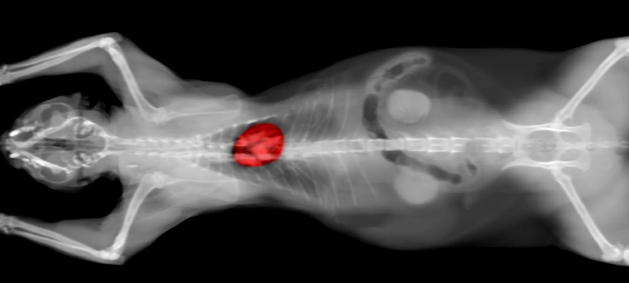
Computed Tomography is a way of examining bodily organs by scanning them with X rays and using a computer to construct a series of cross-sectional images that run along a single plane.

During the test, the patient lays on a table that is attached to the CT scanner. The CT scanner is a very large doughnut-shaped machine with a hole large enough for the table the patient is laying on to pass through. The CT scanner sends X-rays through the area of the body that is being studied. Each rotation of the scanner produces a thin photo slice of the area. All of the pictures are then put together in the computer to create a complete image.
In some cases, a dye called contrast may be used by injecting it in to the bloodstream or joints to see those areas better. For some types of CT scans, the patient may ingest the dye.
A Computed Tomography Scan can be used on nearly any part of the body including the heart, lungs, chest, belly, pelvis, arms and legs. It can take pictures of bodily organs, such as the liver, pancreas, intestines, kidneys, bladder and glands. It also can study cardiovascular systems, bones, and the spinal cord.
Wondering what the difference in a computed tomography (CT) scan and magnetic imaging (MRI) is?
A CT Scan is commonly used for viewing bone injuries, diagnosing lung and chest problems, and detecting cancers. An MRI is well suited for examining soft tissue in ligament and tendon injuries, spinal cord injuries, brain tumors, etc. CT scans are widely used in emergency rooms because the scan takes fewer than 5 minutes. An MRI, can sometimes take up to 30 minutes per study. Often, a veterinarian will need to make multiple studies.
As a rule, an MRI costs more than a CT scan. One advantage of Magnetic Resonance Imaging is that it does not use radiation while Computed Tomography does.
