Animal Specialty & Emergency Center of Brevard
Magnetic Resonance Imaging (MRI)
Magnetic Resonance Imaging provides good contrast between different tissues of the body.

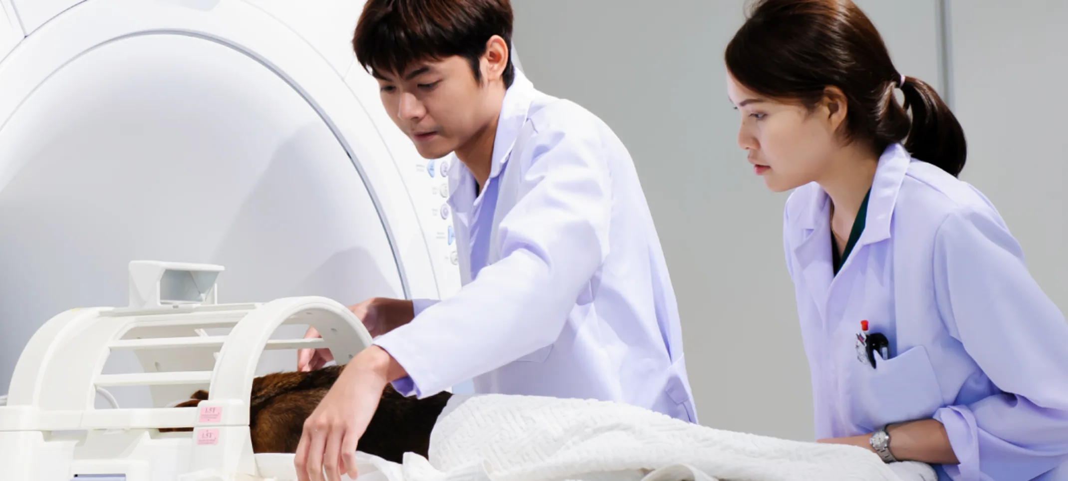
Magnetic resonance imaging (MRI) is a test that uses a magnetic field and pulses of radio wave energy to make pictures of organs and structures inside the body. In many cases MRI gives different information about structures in the body than can be seen with an X-ray, ultrasound, or computed tomography (CT) scan.
For an MRI test, the area of the body being studied is placed inside a special machine that contains a strong magnet. Pictures from an MRI scan are digital images that can be saved and stored on a computer for more study. The images also can be reviewed remotely, such as in a clinic or an operating room.
It is used to find problems such as tumors, bleeding, injury, blood vessel diseases, or infection. MRI also may be done to provide more information about a problem seen on an X-ray, ultrasound scan, or CT scan. Contrast material may be used during MRI to show abnormal tissue more clearly.
Wondering what the difference in a Computed Tomography (CT) Scan and Magnetic Resonance Imaging (MRI) is?
A CT Scan is commonly used for viewing bone injuries, diagnosing lung and chest problems, and detecting cancers. An MRI is well suited for examining soft tissue in ligament and tendon injuries, spinal cord injuries, brain tumors, etc. CT scans are widely used in emergency rooms because the scan takes fewer than 5 minutes. An MRI, can sometimes take up to 30 minutes per study. Often, a veterinarian will need to make multiple studies.
As a rule, an MRI costs more than a CT scan. One advantage of Magnetic Resonance Imaging is that it does not use radiation while Computed Tomography does.
