Animal Specialty & Emergency Center of Brevard

Cancer-Related Surgery
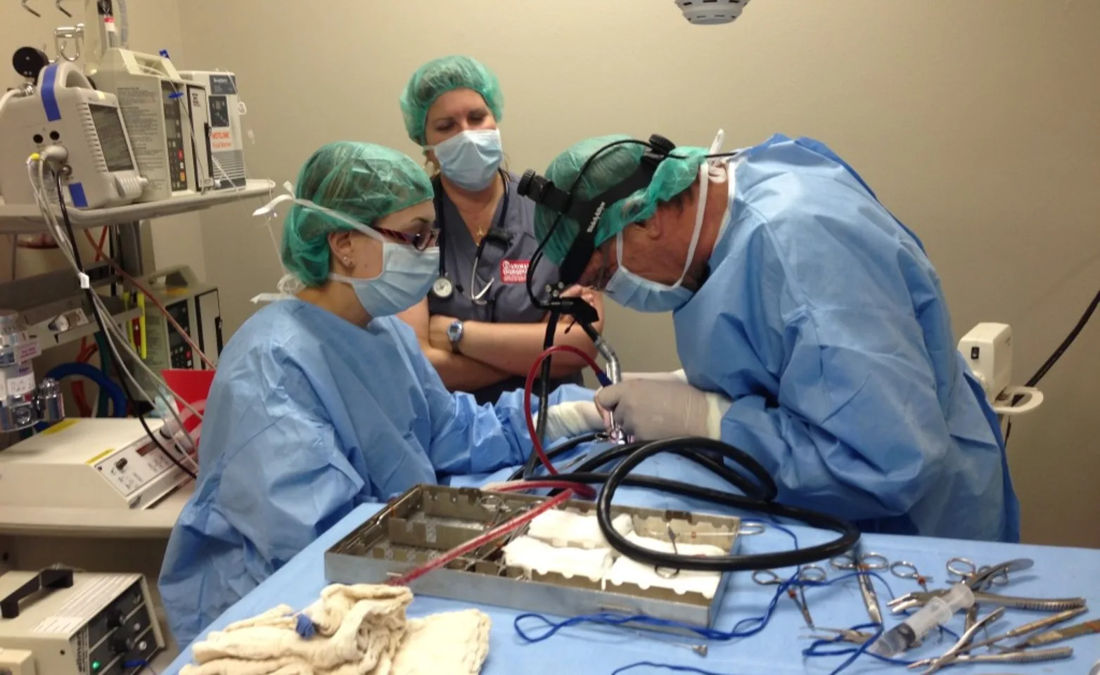
With many types of animal cancer, surgery is the primary recommended treatment, and the best chance for cure or long-term resolution is with a definitive first surgery. Surgery also allows a tissue diagnosis to best determine the extent of therapy required.
Advanced Imaging greatly aids the pre-operative staging and surgical planning so that large and/or invasive tumors can be safely and appropriately removed, both minimizing recurrence and maximizing your pet’s post-operative comfort and function.

When appropriate, some tumors may be removed via minimally invasive surgery. In some urethral or colonic cancer management, stents may be placed within collapsed or obstructed structures. Hyperbaric Oxygen Therapy may also help improve your pet’s post-operative cancer survival rate.
Many pets may experience long-term resolution or even cure, gaining extended periods of quality of life where they have little or no signs of disease. Depending on the diagnosis, follow up therapies may or may not be indicated. These therapies include chemotherapy (which may be performed in-house by our internal medicine specialist, Dr. Levine) and radiation therapy, for which we may refer you. A team and multi-disciplinary approach is best to prevent local recurrence and distant spread.
Congenital Defects Surgery
Portosystemic shunts – a vessel that causes blood to bypass the liver. These can be diagnosed with bloodwork, identified with surgery, confirmed with an intra-operative x-ray study, and treated by using an Ameroid Constrictor Ring, which allows the shunt to close slowly to minimize complications.
Patent Ductus Arteriosus (PDA) – a vessel between the aorta (the main artery of the heart) and pulmonary artery (supplying the lungs) that can cause reduced growth, damage to the heart, and often death within a year due to heart failure. This can be surgically closed and potentially give a patient a normal life.
Thoracic Surgery
Some of these may be accomplished through minimally invasive surgery:
Patent Ductus Arteriosus (PDA)
Pericardiectomy – removal of the sac around the heart, which can become distended with fluid and compress the heart, causing congestive heart failure
Thoracic duct ligation – for chylothorax (accumulation of lymph fluid in the chest)
Lung lobectomy – for lung torsion, tumor, bulla (air bubbles that can rupture and cause air in the chest to compress the lungs), or severe bacterial or fungal infection
Resection of tumors on certain parts of the heart, such as removal of hemangiosarcoma from the right atrium
Esophageal obstruction not amenable to endoscopic removal
Hernia Repair
Defects in muscle walls which may be congenital, developmental or traumatic in origin.
Synthetic mesh may be used in some instances.
Minimally Invasive Surgery
Laparoscopy and Thoracoscopy allow viewing of highly detailed camera images of internal organs in the abdomen and chest via small portals, rather than large incisions. Biopsies can be obtained via these portals, and certain procedures can be performed, such as pericardial window (to treat fluid within the sac around the heart, which can cause congestive heart failure), tumor exteriorization and removal.
Endoscopic-assisted Gastropexy – For prevention of Gastric Dilation-Volvulus (GDV; commonly referred to as “bloat,” but with a life-threatening twist in the stomach). An endoscope is passed down the throat into the stomach, which is then inflated and illuminated. Via this means, the stomach can be sutured to the body wall through a 4 cm (less than 2 inch) incision, preventing this life-threatening problem from occurring.
STENT Placement – via fluoroscopy or endoscopy, stents can be placed within collapsed or obstructed (due to scar tissue, tumor, etc.) structures, such as the trachea (windpipe), urethra, ureters (the tubes carrying urine from the kidney to the bladder), and even colon. Stents are sometimes used for urethral and colonic cancer management. Surgery may not be required at all.
